Why are oxygen levels in brains of MS patients unusually low?

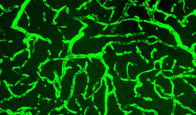
A photograph of blood vessels in the brain of an animal model of MS. They are green in this image because a green fluorescent dye was added, making capillaries visible under a microscope.

Yang, R. and J. F. Dunn (2015) Reduced cortical microvascular oxygenation in multiple sclerosis: a blinded, case-controlled study using a novel quantitative near-infrared spectroscopy method. Sci Rep 5: 16477.

link to paper here (open access)


Runze and I just published this paper. It has two major new findings. One is that, in about ½ of patents with MS, the brain shows signs of hypoxia (unusually low oxygen levels). This is based on quantifying the amount of oxygen bound to hemoglobin in the microvessels. The other news is that we could obtain a measure of brain oxygen using a method that is easily translatable to a clinic, doesn’t hurt and only takes a minute. It is less inconvenient than having your blood pressure measured in your arm. As we move forward, we have to find out why this hypoxia exists—could it be a totally new measure of the amount of inflammation in brain? I’m sure it will tell us something new and may become a new method for detecting treatment response.

In my opinion, this paper opens up a totally new way of looking at MS—much like MRI did when they first showed that plaques existed. We are taking steps towards understanding what is changing in the brain of patients with MS.

Let me explain.

Hypoxia is a term for low oxygen. If you climb a mountain, there is less oxygen and your body is hypoxic relative to a person living at sea-level. Different methods are used to measure oxygen in brain because a direct measurement is pretty difficult—unless you implant a probe to quantify oxygen. In our study, we used a method called near-infrared spectroscopy or NIRS.

Hemoglobin is a protein in the blood that carries oxygen. If all hemoglobin has oxygen bound, then the oxygen saturation is 100%. Light is absorbed differently if the hemoglobin is bound to oxygen or not. That is why blood in arteries is red and blood in veins is more purple or bluish in color. With NIRS, we shine light into the head and quantify the amount of oxygen bound to hemoglobin, by looking at the different color of light coming to our detector. The method focuses on the small vessels, like the ones in the image above, where oxygen is being transferred into the brain (the capillaries and microvessels). Most NIRS can’t quantify between subjects. Our method uses frequency domain NIRS to obtain absolute values. Big words, but the main point is that we can get an absolute measurement of the oxygen saturation of hemoglobin.

We found that about ½ of patients with MS were hypoxic—using the definition that the amount of oxygen bound to hemoglobin was lower than in control subjects. This wasn’t caused by low inspired oxygen, or an inability of the lungs to work. The low oxygen related to a low delivery of oxygen to the brain relative to how much is being used. A kind of supply problem.

It is too early to say if this hypoxia is damaging to the brain. The brain is pretty tolerant of low oxygen. It is very likely that the hypoxia will change how the immune response functions in the brain. Hypoxia could make the immune response worse.

A second large benefit to our paper is we show that our NIRS method is capable of detecting the hypoxia. The method is easy to use and only takes a few minutes of your time. It involves holding a small pad against the forehead for a minute.

Now that we have shown that hypoxia exists, and that we can measure it, the next chapter begins. Why is it there? Is it a marker of unregulated inflammation? Could we use it to tell if anti-inflammatory drugs are working? Inflammation can damage the blood vessels in a way that changes how flow is regulated. Cells responsible for Inflammation can also use a lot of oxygen. Should we re-investigate oxygen therapies? Could it be that major oxygen therapy trials didn’t show significant results because they only worked in the patients that were already hypoxic? At the time of the trials, we had no way to tell, before treatment, if the brain was hypoxic or not so all patients were treated equally.

We have a new method and are now showing that it is sensitive to changes in the brain of patients with MS.

Another cool part of this study is that the funding to do it came from undergraduate student salary grants. This was not a big, well funded study. We cobbled together money for Runze (we wrote this while he was an undergraduate student). Most of his funding was from Alberta Innovates Health Solutions. The equipment funding came from AIHS and Canada Foundation for Innovation.


The skiers guide to growing your own heel spur

An earlier version was Published in the Fortress Alpine Ski Team newsletter


An earlier version was Published in the Fortress Alpine Ski Team newsletter

Our adventure into heel spur growth began last year when our son was 12, and skiing every moment he could. There was a two week period where they were racing 3 days each week and adding training on top. Everyone was exhausted—too much tuning, being cold, sore muscles and general fatigue. At the end of this, “Manchild” pulled his socks off to show his coach his sore heels and, “ voila”, he had large growths on the back of both heels, just outside of the centre line. They were over a cm deep, large enough to name, angry red and very sore.

So what happened, what is it, how can you prevent it and what can you do about it? Depending on the location on your foot, these bumps can mean different things. The ones at the back of the heel, like my son Robert’s little friends, are at the insertion of the Achilles tendon and the bone.

Here is the sum of my meager knowledge. The boot fitting community is, to a large extent, sure that the growth is due to poor boot fit. This is usually because they are too tight, but they also might be too loose. The latter can cause your foot to bang against the back of the boot. Good fitters can find the sensitive site and grind a pocket or blow the boot out to relieve the pressure. But hold on—is this all there is?

I dug more into the literature and pestered my colleagues at the University of Calgary. Their input is unofficial—in that I nabbed them by email or in the halls, and launched into my characteristic gambit “This is really interesting what do you think?” You see, I don’t totally agree with the bootfitters. If it was just mechanical, why was there no other bruising? If something is growing, the body has to also grow new blood vessels to feed the bump, and that usually involves inflammation. If the tendon is inflamed, can the bump also be caused by Achilles tendon injury?

Dr. Benno Nigg is the Director of the Human Performance Laboratory at the University of Calgary. He is also a good neighbor, although he feeds the deer– much to the consternation of the gardeners in the neighborhood. He has worked with Nike and Adidas on high performance shoe design, and has had contracts with the national ski team concerning boot fitting. Benno thought the tendon injury theory had some merit, and thought that pre-exercise active stretching of the tendon may help a lot to prevent further injury. What the heck, good stretching won’t usually hurt so it’s worth a try.

Dr. Preston Wiley is a sports medicine physician in interested in “overuse injuries” in the Roger Jackson Sports Medicine centre at the UofC. He had a PhD student working on reducing pain associated with the bone spur. When I told him Robert was 12 when it happened he exclaimed—“Sever’s disease, it’s relatively common when boys are 12 and girls are 11”. http://kidshealth.org/parent/medical/bones/severs_disease.html# .

Sever’s disease is an acute injury to the growth plate at the site of insertion of the Achilles tendon. It comes on quickly, it will generate a bump, and the pain will go away in 1-2 years as the site heals up. Older athletes get bumps in the same area, but these are called “pump bumps” which alludes to the fact that ladies in high heels get them too. This is more what the boot fitters are thinking of, in that it’s caused by a combination of tension on the tendon and rubbing of the insertion. So the bump might be from rubbing, but keep in mind it may be a growth plate injury, and in part it relates to tendon injury. Also, it isn’t usually bone. It’s usually scarring of the tendon.

So, the bump can be caused by more than one thing. I suggest that f there is acute pain when you are young, try to see a sports medicine physician. The standard pump bump is only alleviated by VERY active icing after your sport and reducing the rubbing. Get on the problem early and fast. Working through the pain in this case will only cause you more problems. Work on the boot fitting. Once the bump hardens and calcifies, there may not be much you can do about it. From our experience, naming them at least makes it more fun to have them.