Are the wires in the brain damaged after concussion?


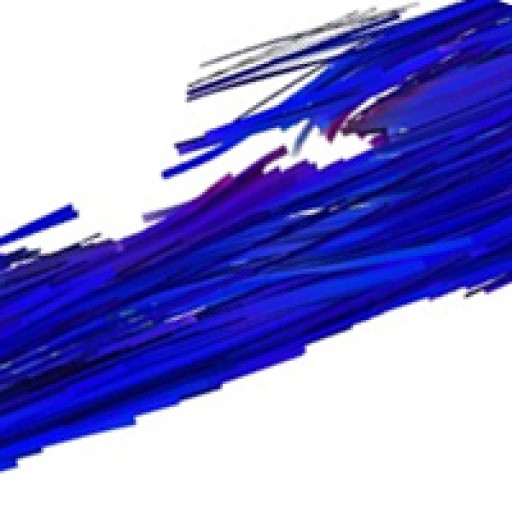
cropped-mouse-spinal-cord-DTI-600-1.jpgDiffusion tensor MRI showing breaks on long fibre tracts when myelin is lost. This example is from a model of multiple sclerosis. See explanation below. Thanks to the team at the Experimental Imaging Centre and Dayae Jeong.


What is injured in your brain after a concussion? It is pretty obvious that something isn’t quite right. There is a list of symptoms in the sport concussion assessment tool three, the SCAT3, that you can look at for a range of symptoms.

http://bjsm.bmj.com/content/47/5/259.full.pdf

The brain has long communicating fibre bundles called nerve tracts. These are clearly visible on MRI. The problem that MRI researchers have had in the past is that the changes are very subtle and not visible on standard MRI’s.

To overcome this problem, MRI groups have been working intensely to make the images more sensitive to the injury. There are MRI methods, called “sequences” by MRI specialists, which are showing promise as being sensitive to structural injury. These sequences are sensitive to the amount of myelin. Myelin is the covering on nerves that helps the signal travel down the nerve. When you lose myelin, as occurs in conditions such as multiple sclerosis, the nerve doesn’t function as well. If the myelin doesn’t regrow, the nerve may die.

Clinical MRI systems are actually imaging protons and the most abundant source of protons in the body is in water. So MRI is really imaging water. The trick to making contrast in the image is to make the sequence sensitive to things that can change in the water.

For instance, water can diffuse or move. In the brain, water diffuses fairly readily along fibre tracts but does not readily diffuse across a tract. This is because myelin is similar to soap, and it repels water. An MRI sequence called diffusion tensor imaging has emerged which is sensitive to the amount of water and the direction the water is diffusing. If water diffuses randomly in all directions, it is called anisotropic diffusion. If the diffusion has a directionality, it is isotropic. The measure of this is the fractional anisotropy or FA.

If myelin is damaged, water can more readily diffuse in all directions and the FA will decline. Such changes have been observed indicating that myelin may be damaged in certain regions of the brain after mTBI.

Dean, P. J., J. R. Sato, G. Vieira, A. McNamara and A. Sterr Long-term structural changes after mTBI and their relation to post-concussion symptoms. Brain Inj: 1-8.

Shenton, M. E., H. M. Hamoda, J. S. Schneiderman, S. Bouix, O. Pasternak, Y. Rathi, M. A. Vu, M. P. Purohit, K. Helmer, I. Koerte, A. P. Lin, C. F. Westin, R. Kikinis, M. Kubicki, R. A. Stern and R. Zafonte A review of magnetic resonance imaging and diffusion tensor imaging findings in mild traumatic brain injury. Brain Imaging Behav 6(2): 137-92.


Another sequence that looks at myelin is called quantitative T2. A very specific, not widely available variant is called imaging myelin water fraction or MWF. The University of British Columbia MRI group has been a leader in this area. They looked at MWF in athletes and, indeed, found reduced MWF along major fibre tracts in brain. http://journals.plos.org/plosone/article?id=10.1371/journal.pone.0150215

Again, this supports the idea that damage to myelin occurred.


Wright, A. D., M. Jarrett, I. Vavasour, E. Shahinfard, S. Kolind, P. van Donkelaar, J. Taunton, D. Li and A. Rauscher (2016) Myelin Water Fraction Is Transiently Reduced after a Single Mild Traumatic Brain Injury – A Prospective Cohort Study in Collegiate Hockey Players. PLoS One 11(2): e0150215.


Another sequence is called magnetization transfer imaging or MTI. This one looks at how water interacts with surrounding chemicals and how those chemical transfer some of their MRI related characteristics (or magnetization) to water. This is where the label “magnetization transfer” arose. One can design a sequence such that a lot of magnetization is transferred to water if myelin is present, but not much if myelin is not present. So the MT declines as myelin is lost.

There has been less work on MTI, but I expect to see a lot more. What has been done though, is consistent with the other methods. A decline in MT has been observed.


McGowan, J. C., J. H. Yang, R. C. Plotkin, R. I. Grossman, E. M. Umile, K. M. Cecil and L. J. Bagley (2000) Magnetization transfer imaging in the detection of injury associated with mild head trauma. AJNR Am J Neuroradiol 21(5): 875-80.

Narayana, P. A., X. Yu, K. M. Hasan, E. A. Wilde, H. S. Levin, J. V. Hunter, E. R. Miller, V. K. Patel, C. S. Robertson and J. J. McCarthy (2014) Multi-modal MRI of mild traumatic brain injury. Neuroimage Clin 7: 87-97.

So, there are three MRI methods that have been used to show a loss of myelin after concussion or mTBI. This is worrying as it is very difficult to re-grow myelin to its original form. It is encouraging as there are imaging methods on the horizon that may help identify the extent of injury after concussion.

Diagnosing concussion is one thing, and we probably do that pretty well now. However to manage the injury we need to know the magnitude of the injury, where the injury has occurred, and whether the injury has recovered. This is the holy grail of brain imaging in the field of concussion research.

CONCUSSION: Stealing your mind

DSC_1776Dear everyone suffering from concussion, or trying to help someone who has a concussion:

Here is a podcast series on concussion.

CONCUSSION: Stealing your mind

Episode 1 is about concussion–what is it

Concussion is also called mild traumatic brain injury or mTBI. It is a brain injury. The symptoms are extremely variable.

Episode 2 is about diagnosis of concussion.

I interviewed Dr. Karen Barlow from the Alberta Children’s Hospital.

For most people, diagnosis is done with a low bar—by this I mean few things are needed for you as a coach or parent to consider concussion as being likely.   I recommend you download and read a great resource—the statement on concussion in sport (or the SCAT3).

http://www.nrl.com/portals/nrl/RadEditor/Documents/ConcussioninSport.pdf

Consensus Statement on Concussion in Sport—the 4th International Conference on Concussion in Sport Held in Zurich, November 2012 Paul McCrory, MBBS, PhD, Willem Meeuwisse, MD, PhD, Mark Aubry, MD, Bob Cantu, MD, Jiri Dvorak, MD, Ruben J. Echemendia, PhD, Lars Engebretsen, MD, PhD, Karen Johnston, MD, PhD, Jeffrey S. Kutcher, MD, Martin Raftery, MBBS, Allen Sills, MD, and Coauthors: Brian W. Benson, MD, PhD, Gavin A. Davis, MBBS, Richard G. Ellenbogen, MD, Kevin M. Guskiewicz, PhD, ATC, Stanley A. Herring, MD, Grant Iverson, PhD, Barry D. Jordan, MD, MPH, James Kissick MD, CCFP, Dip Sport Med, Michael McCrea, PhD, ABPP, Andrew S McIntosh, MBiomedE, PhD, David L. Maddocks, LLB, PhD, Michael Makdissi, MBBS, PhD, Laura Purcell, MD, FRCPC, Margot Putukian, MD, Michael Turner MBBS, Kathryn Schneider, PT, PhD, Charles H. Tator, MD, PHD


Episode 3 is about concussion management. In this Episode I interviewed Dr. Karen Barlow from the Alberta Children’s Hospital (and Alberta Children’s Hospital Research Institute) and Dr. Chantel Debert from the Foothills hospital (and the Hotchkiss Brain Institute).

I hope this series gives you some good ideas.


I do research into brain—I study new imaging methods and I use those imaging methods to unlock secrets about brain injury and disease. We are working on a large project using light to image brain—a method that can monitor brain activity (functional near-infrared spectroscopy or fNIRS) while sittng comfortably in a chair (or on a bike, a sports facility or medical clinic).

One of my main projects right now involves brain injury and concussion.

@jeffreyfdunn

@imaginer (both the same twitter accounts)

The skiers guide to growing your own heel spur

An earlier version was Published in the Fortress Alpine Ski Team newsletter


An earlier version was Published in the Fortress Alpine Ski Team newsletter

Our adventure into heel spur growth began last year when our son was 12, and skiing every moment he could. There was a two week period where they were racing 3 days each week and adding training on top. Everyone was exhausted—too much tuning, being cold, sore muscles and general fatigue. At the end of this, “Manchild” pulled his socks off to show his coach his sore heels and, “ voila”, he had large growths on the back of both heels, just outside of the centre line. They were over a cm deep, large enough to name, angry red and very sore.

So what happened, what is it, how can you prevent it and what can you do about it? Depending on the location on your foot, these bumps can mean different things. The ones at the back of the heel, like my son Robert’s little friends, are at the insertion of the Achilles tendon and the bone.

Here is the sum of my meager knowledge. The boot fitting community is, to a large extent, sure that the growth is due to poor boot fit. This is usually because they are too tight, but they also might be too loose. The latter can cause your foot to bang against the back of the boot. Good fitters can find the sensitive site and grind a pocket or blow the boot out to relieve the pressure. But hold on—is this all there is?

I dug more into the literature and pestered my colleagues at the University of Calgary. Their input is unofficial—in that I nabbed them by email or in the halls, and launched into my characteristic gambit “This is really interesting what do you think?” You see, I don’t totally agree with the bootfitters. If it was just mechanical, why was there no other bruising? If something is growing, the body has to also grow new blood vessels to feed the bump, and that usually involves inflammation. If the tendon is inflamed, can the bump also be caused by Achilles tendon injury?

Dr. Benno Nigg is the Director of the Human Performance Laboratory at the University of Calgary. He is also a good neighbor, although he feeds the deer– much to the consternation of the gardeners in the neighborhood. He has worked with Nike and Adidas on high performance shoe design, and has had contracts with the national ski team concerning boot fitting. Benno thought the tendon injury theory had some merit, and thought that pre-exercise active stretching of the tendon may help a lot to prevent further injury. What the heck, good stretching won’t usually hurt so it’s worth a try.

Dr. Preston Wiley is a sports medicine physician in interested in “overuse injuries” in the Roger Jackson Sports Medicine centre at the UofC. He had a PhD student working on reducing pain associated with the bone spur. When I told him Robert was 12 when it happened he exclaimed—“Sever’s disease, it’s relatively common when boys are 12 and girls are 11”. http://kidshealth.org/parent/medical/bones/severs_disease.html# .

Sever’s disease is an acute injury to the growth plate at the site of insertion of the Achilles tendon. It comes on quickly, it will generate a bump, and the pain will go away in 1-2 years as the site heals up. Older athletes get bumps in the same area, but these are called “pump bumps” which alludes to the fact that ladies in high heels get them too. This is more what the boot fitters are thinking of, in that it’s caused by a combination of tension on the tendon and rubbing of the insertion. So the bump might be from rubbing, but keep in mind it may be a growth plate injury, and in part it relates to tendon injury. Also, it isn’t usually bone. It’s usually scarring of the tendon.

So, the bump can be caused by more than one thing. I suggest that f there is acute pain when you are young, try to see a sports medicine physician. The standard pump bump is only alleviated by VERY active icing after your sport and reducing the rubbing. Get on the problem early and fast. Working through the pain in this case will only cause you more problems. Work on the boot fitting. Once the bump hardens and calcifies, there may not be much you can do about it. From our experience, naming them at least makes it more fun to have them.