Or using 2020 vision to visualize the future of Magnetic Resonance
Or Magnetic Resonance, from molecules to space science
Are you doing magnetic resonance research (MRI, MRS, EPR)? Are you keen on learning cool new stuff, brainstorming new ideas and networking with peers and leaders in the field?
Come to the Gordon Research Conference and Seminar this summer!
Gordon Research Conference on in vivo Magnetic Resonance: July 19-24 (open to all)
https://www.grc.org/in-vivo-magnetic-resonance-conference/2020/
Chair: Jeff F Dunn Vice Chair: Kim Butts Pauly
Gordon Research Seminar on in vivo Magnetic Resonance July 18-19 (trainees-students/pdocs) Chairs: Manushka Vaidya and Melissa Haskell
https://www.grc.org/in-vivo-magnetic-resonance-grs-conference/2020/
Why come?
This meeting has been very influential in the development of magnetic resonance. It is one of those small meetings, in a beautiful setting, where people can meet others in their field and where ideas flow. This is the 20th anniversary meeting. In recognition of this, the meeting will have a bit of a historical feeling as well as a goal of brainstorming the next 20 years. It will cover many aspects of MRI including hardware, sequences and processing. From the first meeting, chaired by Michael Smith, to the 2018 meeting, chaired by Dan Sodickson, this meeting on Magnetic Resonance has been ranked among the top Gordon Research Conference meetings.
If you go farther back, the current GRC evolved from the GRC on Magnetic Resonance in Biology and Medicine, which started in 1974. The last meeting, in 1998, was chaired by David Wemmer, had Alex Pines as a speaker, and MRI was a minor component.
As a special anniversary theme, we are asking speakers to think about major discoveries in their labs, and to wax profound about where thire field might go in the next 20 years.
What is a Gordon Conference?
My students have said that this is the best conference they have ever gone to. It is one of those small group meetings where ideas flow and friends are made. You will hear news from a range of MR fields—expanding your knowledge and contacts.
Gordon Conferences are a special genre. The Gordon organizes sites, and all the meeting logistics. The program is required to have time for discussion and there are no parallel sessions. Speakers are encouraged to include unpublished date. People often come and don’t present. They are there to learn what’s new and to discuss science with leaders and trainees. One of the unique things about the “Gordon” is that it is a closed meeting. You have to submit an application and obtain approval to register. There is no abstract book. Photographs are not allowed without specific permission. Thus everyone’s intellectual property is protected. This conference is aimed at learning what is happening at the forefront of field. People can, and do, discuss what is new in their lab—not just what is already published. It was the Gordon that hosted many of the early meetings where the idea of MRI was tossed around, and developed.
They GRC is about networking and discussions—and so there is plenty of time in the afternoon and evenings for getting to know other attendees. Our GRC is famous for a nice hill walk, swimming, and the famous soccer (football!) games. The GRC starts with dinner on Sunday, an evening session and meetings in the bar (a relaxing venue where you don’t have to have alcohol to have a good time).
Posters are encouraged so bring it on. These will be relaxed sessions where everyone will have time to meet people at the poster sessions.
What is the Gordon Seminar?
This is a trainee only meeting. It runs from the day before the GRC starts, to the afternoon of the first day of the GRC. Most speakers and discussion leaders are trainees. Get to know your peers.
So—great science, cutting edge AND time to meet people—not just hear them while they blather on behind a podium.
———————————————————————————-
Hints for attending a Gordon Conference/Seminar:
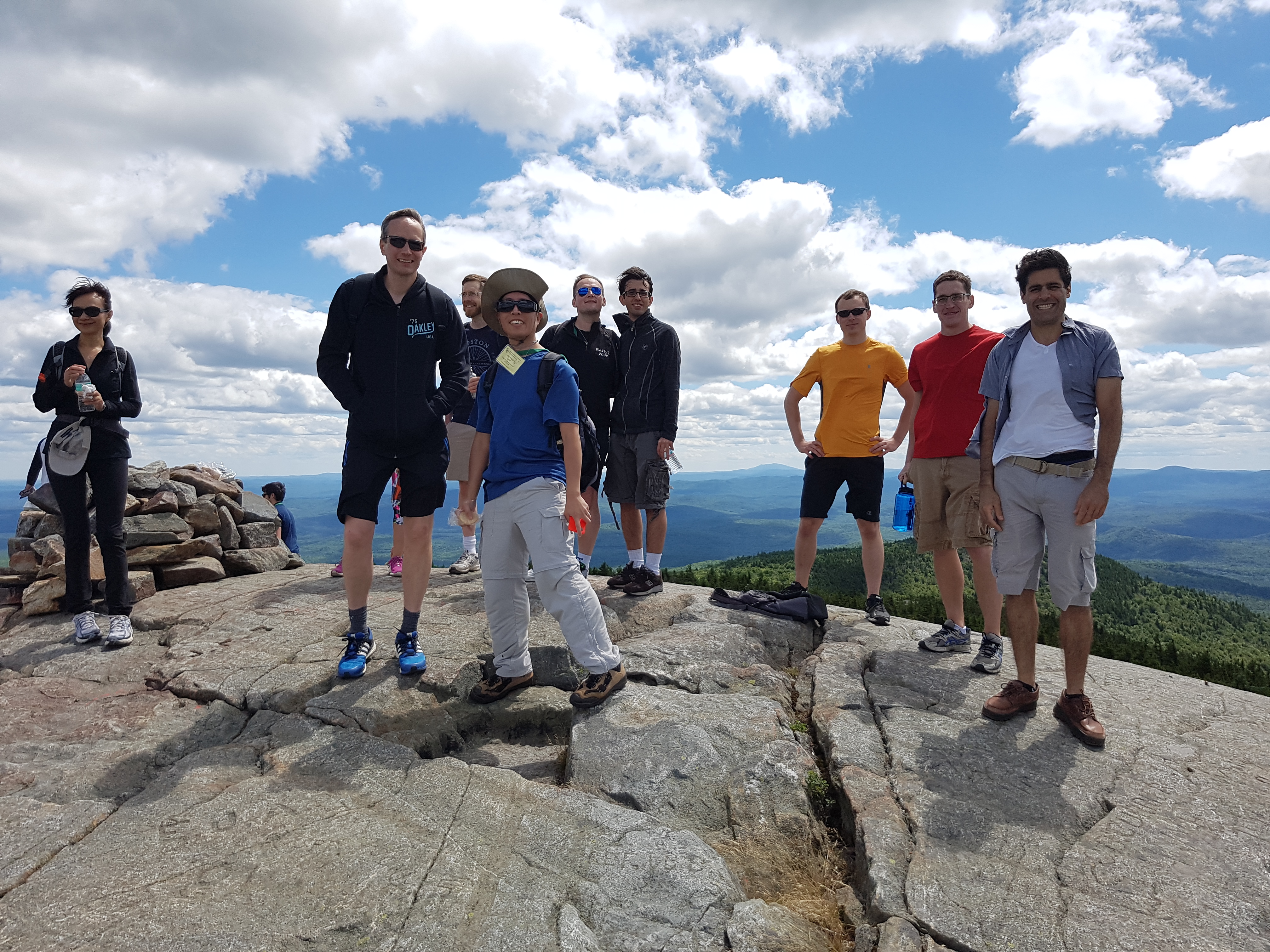
Attendees and speakers are encouraged to stay for the duration so you have time to make friends and go away with new colleagues. There are many late night brainstorming sessions sitting on the lawn with the stars overhead (hope for no rain!). There are organized social events every day including the now famous Kearsarge hike with fantastic views of New England.
Bring walking shoes, a bathing suit for the freshwater lakes and whatever sport gear you might want. Take in organized activities such as canoeing. Partake of the famous “last night” Gordon dinner of lobster (have I mentioned that the food is fantastic and the bar is cheap).
To register follow the links above for the GRC and GRS. Remember that it is a two step process where you apply, obtain permission and then register. We are looking for funding to help people with registrations and note that there is a diversity funding link on the conference homepage.
We look forward to meeting you.
Yours in Magnetic Resonance
Jeff F. Dunn Chair GRC
Kim Butts Pauly Vice-Chair GRC
Manushka Vaidya Co-Chair GRS Melissa Haskell Co-C