Are the wires in the brain damaged after concussion?


cropped-mouse-spinal-cord-DTI-600-1.jpg
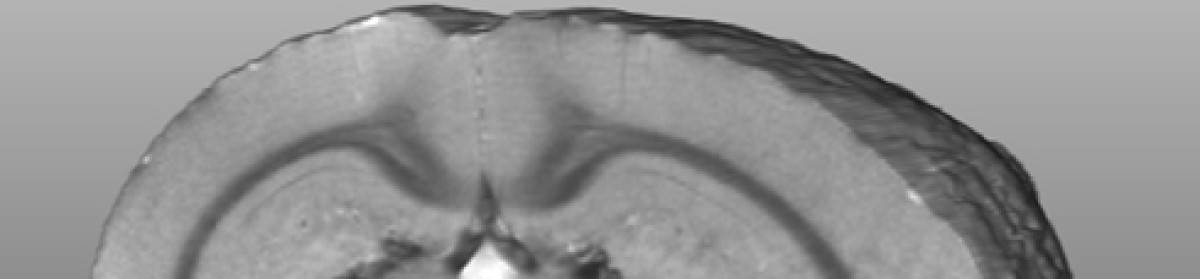
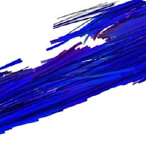
Diffusion tensor MRI showing breaks on long fibre tracts when myelin is lost. This example is from a model of multiple sclerosis. See explanation below. Thanks to the team at the Experimental Imaging Centre and Dayae Jeong.


What is injured in your brain after a concussion? It is pretty obvious that something isn’t quite right. There is a list of symptoms in the sport concussion assessment tool three, the SCAT3, that you can look at for a range of symptoms.

http://bjsm.bmj.com/content/47/5/259.full.pdf

The brain has long communicating fibre bundles called nerve tracts. These are clearly visible on MRI. The problem that MRI researchers have had in the past is that the changes are very subtle and not visible on standard MRI’s.

To overcome this problem, MRI groups have been working intensely to make the images more sensitive to the injury. There are MRI methods, called “sequences” by MRI specialists, which are showing promise as being sensitive to structural injury. These sequences are sensitive to the amount of myelin. Myelin is the covering on nerves that helps the signal travel down the nerve. When you lose myelin, as occurs in conditions such as multiple sclerosis, the nerve doesn’t function as well. If the myelin doesn’t regrow, the nerve may die.

Clinical MRI systems are actually imaging protons and the most abundant source of protons in the body is in water. So MRI is really imaging water. The trick to making contrast in the image is to make the sequence sensitive to things that can change in the water.

For instance, water can diffuse or move. In the brain, water diffuses fairly readily along fibre tracts but does not readily diffuse across a tract. This is because myelin is similar to soap, and it repels water. An MRI sequence called diffusion tensor imaging has emerged which is sensitive to the amount of water and the direction the water is diffusing. If water diffuses randomly in all directions, it is called anisotropic diffusion. If the diffusion has a directionality, it is isotropic. The measure of this is the fractional anisotropy or FA.

If myelin is damaged, water can more readily diffuse in all directions and the FA will decline. Such changes have been observed indicating that myelin may be damaged in certain regions of the brain after mTBI.

Dean, P. J., J. R. Sato, G. Vieira, A. McNamara and A. Sterr Long-term structural changes after mTBI and their relation to post-concussion symptoms. Brain Inj: 1-8.

Shenton, M. E., H. M. Hamoda, J. S. Schneiderman, S. Bouix, O. Pasternak, Y. Rathi, M. A. Vu, M. P. Purohit, K. Helmer, I. Koerte, A. P. Lin, C. F. Westin, R. Kikinis, M. Kubicki, R. A. Stern and R. Zafonte A review of magnetic resonance imaging and diffusion tensor imaging findings in mild traumatic brain injury. Brain Imaging Behav 6(2): 137-92.


Another sequence that looks at myelin is called quantitative T2. A very specific, not widely available variant is called imaging myelin water fraction or MWF. The University of British Columbia MRI group has been a leader in this area. They looked at MWF in athletes and, indeed, found reduced MWF along major fibre tracts in brain. http://journals.plos.org/plosone/article?id=10.1371/journal.pone.0150215

Again, this supports the idea that damage to myelin occurred.


Wright, A. D., M. Jarrett, I. Vavasour, E. Shahinfard, S. Kolind, P. van Donkelaar, J. Taunton, D. Li and A. Rauscher (2016) Myelin Water Fraction Is Transiently Reduced after a Single Mild Traumatic Brain Injury – A Prospective Cohort Study in Collegiate Hockey Players. PLoS One 11(2): e0150215.


Another sequence is called magnetization transfer imaging or MTI. This one looks at how water interacts with surrounding chemicals and how those chemical transfer some of their MRI related characteristics (or magnetization) to water. This is where the label “magnetization transfer” arose. One can design a sequence such that a lot of magnetization is transferred to water if myelin is present, but not much if myelin is not present. So the MT declines as myelin is lost.

There has been less work on MTI, but I expect to see a lot more. What has been done though, is consistent with the other methods. A decline in MT has been observed.


McGowan, J. C., J. H. Yang, R. C. Plotkin, R. I. Grossman, E. M. Umile, K. M. Cecil and L. J. Bagley (2000) Magnetization transfer imaging in the detection of injury associated with mild head trauma. AJNR Am J Neuroradiol 21(5): 875-80.

Narayana, P. A., X. Yu, K. M. Hasan, E. A. Wilde, H. S. Levin, J. V. Hunter, E. R. Miller, V. K. Patel, C. S. Robertson and J. J. McCarthy (2014) Multi-modal MRI of mild traumatic brain injury. Neuroimage Clin 7: 87-97.

So, there are three MRI methods that have been used to show a loss of myelin after concussion or mTBI. This is worrying as it is very difficult to re-grow myelin to its original form. It is encouraging as there are imaging methods on the horizon that may help identify the extent of injury after concussion.

Diagnosing concussion is one thing, and we probably do that pretty well now. However to manage the injury we need to know the magnitude of the injury, where the injury has occurred, and whether the injury has recovered. This is the holy grail of brain imaging in the field of concussion research.

CONCUSSION: Stealing your mind

DSC_1776Dear everyone suffering from concussion, or trying to help someone who has a concussion:

Here is a podcast series on concussion.

CONCUSSION: Stealing your mind

Episode 1 is about concussion–what is it

Concussion is also called mild traumatic brain injury or mTBI. It is a brain injury. The symptoms are extremely variable.

Episode 2 is about diagnosis of concussion.

I interviewed Dr. Karen Barlow from the Alberta Children’s Hospital.

For most people, diagnosis is done with a low bar—by this I mean few things are needed for you as a coach or parent to consider concussion as being likely.   I recommend you download and read a great resource—the statement on concussion in sport (or the SCAT3).

http://www.nrl.com/portals/nrl/RadEditor/Documents/ConcussioninSport.pdf

Consensus Statement on Concussion in Sport—the 4th International Conference on Concussion in Sport Held in Zurich, November 2012 Paul McCrory, MBBS, PhD, Willem Meeuwisse, MD, PhD, Mark Aubry, MD, Bob Cantu, MD, Jiri Dvorak, MD, Ruben J. Echemendia, PhD, Lars Engebretsen, MD, PhD, Karen Johnston, MD, PhD, Jeffrey S. Kutcher, MD, Martin Raftery, MBBS, Allen Sills, MD, and Coauthors: Brian W. Benson, MD, PhD, Gavin A. Davis, MBBS, Richard G. Ellenbogen, MD, Kevin M. Guskiewicz, PhD, ATC, Stanley A. Herring, MD, Grant Iverson, PhD, Barry D. Jordan, MD, MPH, James Kissick MD, CCFP, Dip Sport Med, Michael McCrea, PhD, ABPP, Andrew S McIntosh, MBiomedE, PhD, David L. Maddocks, LLB, PhD, Michael Makdissi, MBBS, PhD, Laura Purcell, MD, FRCPC, Margot Putukian, MD, Michael Turner MBBS, Kathryn Schneider, PT, PhD, Charles H. Tator, MD, PHD


Episode 3 is about concussion management. In this Episode I interviewed Dr. Karen Barlow from the Alberta Children’s Hospital (and Alberta Children’s Hospital Research Institute) and Dr. Chantel Debert from the Foothills hospital (and the Hotchkiss Brain Institute).

I hope this series gives you some good ideas.


I do research into brain—I study new imaging methods and I use those imaging methods to unlock secrets about brain injury and disease. We are working on a large project using light to image brain—a method that can monitor brain activity (functional near-infrared spectroscopy or fNIRS) while sittng comfortably in a chair (or on a bike, a sports facility or medical clinic).

One of my main projects right now involves brain injury and concussion.

@jeffreyfdunn

@imaginer (both the same twitter accounts)

Concussion– thoughts and suggestions

Have you wondered if you have a concussion? Have you looked on in shock as your child was hit or crashed? Maybe they stopped moving (or worse, started twitching). Brain injury is a huge problem and so I got some of my colleagues together to discuss this in a public forum at Winsport in Calgary.

First in December 2011, and then twice more, I organized an evening to discuss concussion with the sliding sports at Canada Olympic Park. COP kindly provided a venue. Alberta Alpine and COP helped me advertise the event.

I was REALLY fortunate to have two fabulous guest speakers. Dr. Karen Barlow is an Associate Professor of Pediatric Neurology at the Alberta Children’s Hospital Research Institute for Child and Maternal Health. She is a specialist in concussion and traumatic brain injury. Dr. Kelly Brett is a sports medicine physician at the University of Calgary Sports Medicine clinic and is the Head physician for the Calgary Flames hockey team.

The following is my summary of the discussion. I tried to capture the gist of the talks and the discussion.

What is concussion, how do you prevent it, and how do you cure it. If this were all known of course, then there wouldn’t be a problem.

So, what is concussion? Sorry, we don’t know. This is a complex injury.

Concussion is currently defined by the symptoms. There are forms that you can use such as the standard concussion assessment tool (SCAT) or the longer more modern SCAT2 (among others). The biological link, or the precise damage that occurs in the brain that causes these symptoms, just isn’t known.

One can make an educated guess. “Ringing your bell” could break the fine connections that transfer information in the brain, These connections, (the synapses) as well as the wires (the axons), are very fragile. The barrier between the blood and the tissue of the brain, the “blood-brain barrier” is also fragile. The barrier is partly physical, in that there are collections of cells that form a wall between the blood and the brain. The barrier is also “regulatory”, in that transport processes in these barrier cells determine what crosses into the brain or returns to the blood. There can be disruption to either, or both, of the physical and regulatory components of the BBB. Such disruption can cause chemicals to cross into the brain that can damage cells and can cause the brain to swell. There may be metabolic disruption. The energy factory in the cell, the mitochondria, may be damaged in some way.

Headache, nausea, and not feeling quite right are just some of the symptoms. Even “not feeling quite right” should be taken seriously. Keep in mind the headache only comes from the surface of the brain. There are no pain sensors in the brain. Only from surface layer, largely in the membrane called the meninges, do you receive pain signals. So—you can easily see that damage can occur in the brain with no associated pain (or headache).

It’s easy to say you can prevent concussion by removing head hits from sport, but in reality head hits are not the only problem. Your head can get shaken up badly with a hit to the chest or a fall. The head has to move violently to have the brain shaken up. A sharp rap to the head may not cause a concussion due to the resistance in your fluid filled brain box (the skull) that prevents a very sharp knock from moving the brain. Hits or falls which cause large movements of the whole head are usually more serious.

So, we don’t know what it is and we can’t prevent them. We can reduce them through training and good helmets. A helmet won’t eliminate concussions either—sorry. Even with a helmet you can get a whiplash type of injury. We are left with making sure we recognize concussion and that one works hard to minimize the long term damage.

In the old days, two weeks off was considered enough. Now it is understood that each person is an individual and each concussion is unique. There are no generalizations on how long it will take to return to play.

What can be said is that the first hours may be associated with more life threatening problems like bleeding on the brain. It is very important that the subject be properly evaluated. Depending on the symptoms, you may or may not have CT or MRI to look for complications. These imaging methods are used to rule out more severe problems. There is no imaging method currently available that can be used to diagnose concussion itself. MRI holds promise. I’m an MRI researcher so I’m hoping for a home run here.

There are other methods we are working on as well as MRI. If anyone wants to fund a graduate student for my lab, we could work faster on this (just a thought).

After a couple of weeks (kind of like the time needed to heal any other type of bruise), you can start getting serious about return to play. If the person is symptom free, then you can consider doing some exercise during this time. Do some exercise to elevate the heart rate (say to 120). If there are no symptoms, then over the next days gradually increase the exercise. If symptoms occur, back off to the point where there were no symptoms and starts again. If you have had a bad concussion, having medical advice during this phase would be important.

There are advantages to taking concussion seriously. If you “aren’t feeling quite right” and try to throw yourself down a super-G course at 110kph, the results may not be pretty. In other words, your coordination may not be perfect and so you have an increased risk of falling and getting another concussion. A second more obvious reason is that if the damage isn’t healed, you may make it worse.

Some points:

-the force of a hit doesn’t relate well to the extent of injury. You can be hit hard and not have symptoms. You can be hit less violently and have a poor outcome. It may relate to the exact forces translated to the head and the angle of the hit

-you don’t have to go unconscious to have a concussion

-mouth guards and helmets to not prevent concussion

Dr. Barlow noted that most kids get better pretty rapidly, and have no long term symptoms. Dr. Brett noted that if the symptoms aren’t getting better, make sure you have your neck examined. If symptoms have not resolved after a couple of weeks you should see a professional. Keep in mind that upper neck injuries can cause the same symptoms as concussion.

On June 14, 2012, I had the pleasure of listening to Dr. Willem Meeuwisse. He is the Editor in Chief of the Clinical Journal of Sports Medicine. He is a concussion specialist, advises the NHL on concussion and was a coauthor on a recent publication about concussion in the NHL. This paper was linked on the Brain Injury Association of Canada. http://biac-aclc.ca/en/wp-content/uploads/2011/07/NHL_Concussion_CMAJ_2011.pdf

He reiterated much of what I’ve listed in this blog.

It’s now 2015. One of my goals as a medical researcher is to improve the diagnosis and monitoring of concussion patients. I hope that someday soon, there will be more specific answers to some of the three main questions.